


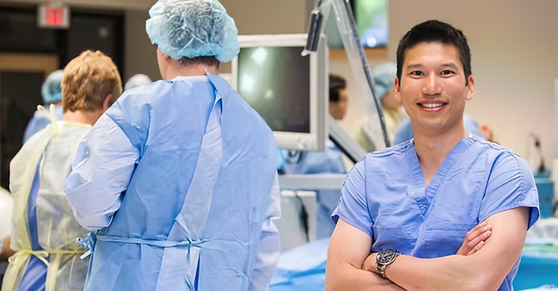
Arthritis, to some, is an inevitable part of the aging process. One doctor in Halifax is working to develop a game-changing piece of technology for both those whose arthritis has left them needing surgery, and the surgeons looking to help alleviate their pain.
Dr. Ivan Wong, a sports medicine and orthopaedic surgeon, and his team are building a 3D ultrasound to allow surgeons to better plan for hip and joint surgeries—to better understand the rigidness of the ball-and-socket joints they are dealing with.
In April 2026, Dr. Wong brought his research and development of this 3D ultrasound to the QEII Foundation’s Innovation Den stage. In this Q&A, Dr. Wong spotlights how a “simple” bedside ultrasound can have a huge impact on Nova Scotia’s healthcare system.
Q: First, can you explain what patients in our healthcare system with hip arthritis may be experiencing when it comes to pain and treatment?
A: When someone has arthritis in their hip joint, it’s due to the deterioration of the protective cartilage in the ball-and-socket hip joint. As this cartilage wears away, the bones rub together, causing chronic inflammation, pain and stiffness. Currently, our healthcare system treats surgery as a last resort. We wait for the joints to fail before we offer the solution, but pain doesn't just stand still, and that pain can lead to other health conditions. But my team felt we could do more for our patients.
Q: Tell us about this bedside 3D ultrasound which you have your team have created.
A: We have been working with Nova Scotia engineers to transform the standard ultrasound, the same technology that you use to look at babies in a womb, into a 3D precision mapping tool for hip conditions. It’s like taking a flashlight into that dark room and tracing the joint with your hand. We combined ultrasound, motion tracking, and artificial intelligence to capture every curving contour of that hip and build a real-time 3D model of each patient's unique joint. Because it is portable, it’s going to be something we can have available right there in a clinic, anywhere. It allows us to get CT level detail without the waiting and without the radiation. It will allow us to diagnose earlier, plan better and intervene before it's too late, hopefully avoiding a hip replacement altogether.
Q: What do you and your fellow surgeons do now if you want a 3D map of the hip area?
A: Right now, the only way to get a 3D map of the hip is by a CT scan, which can come with long wait lists. The average wait is 336 days, so that's nearly another year a patient is losing ground to irreversible arthritis. The cost of this isn't just time, it's safety. CT scanners rely on radiation, and sometimes people need multiple scans. That exposure adds up, and with that, cancer risk goes up.
Q: Is there a surgical option for before you get to a full hip replacement?
A: Yes. It's called hip arthroscopy. Instead of a major operation, a tiny camera goes through small incisions that let us go in and reshape the bone, to stop that sandpaper grinding before the damage is permanent. The goal is by preserving the joint now, we can avoid a replacement later.
Q: What is the next step in your work to make this portable 3D ultrasound come to fruition?
A: My team and I are working to bring this technology to patients. We are hoping to launch the world’s first clinical trial right here at the QEII, giving our patients and communities first access to what I believe will be a new global standard in the race against arthritis. The biggest barrier now is funding the clinical trial so we can prove how well this technology works and ultimately bring it into everyday patient care. With the right support, we can get this trial off the ground, generate the evidence we need, and move this technology from an innovation in the lab to a tool that can benefit patients around the world.
Q: What are some of your hopes for this technology?
A: Sometimes the imaging we do in preparation for a surgery takes so long that the joint looks different by the time we get in there. Imagine being able to show a patient immediately what's going on and starting their intervention before they walk out the door. I hope that this technology will give us the gift of time to make the right care decisions for and with our patients. Ideally, this ultrasound will be at any bedside, at your physician, family doctor or physiotherapist. It can be everywhere to remove more barriers to seeing the right surgeon at the right time and potentially delaying or preventing arthritis from developing.