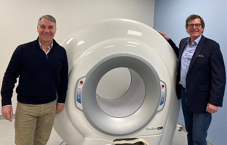
Dr. Jens Heidenreich, head, neuroradiology & head and neck sections at the QEII’s Department of Diagnostic Imaging (left) and Dr. David P. Morris, QEII neurotologist, show off the new cone beam CT scanner at the QEII. The QEII Foundation continues to raise $430,000 to support the acquisition of this first-in-Atlantic Canada technology. CONTRIBUTED
Donor-funded cone beam CT scanner will enhance patient care
For more than half a century the computed tomography scanner — or CT scanner — has been an invaluable tool for obtaining detailed internal images of the human body. This diagnostic tool uses X-rays to create three-dimensional, cross-sectional images that are used to detect a wide range of conditions including tumours, internal bleeding and broken bones, and to guide procedures like surgery and biopsies.
Thanks to QEII Foundation donors, in early October 2025, the first scans using a new, specialized scanner were taken at the Dickson Building at the QEII Health Sciences Centre. The first cone beam CT scanner in Atlantic Canada uses a cone-shaped beam of X-ray light to generate highly detailed images.
“We’re looking at images measured in a fraction of a millimetre, with extremely high resolution,” says Dr. Jens Heidenreich, head of neuroradiology and head and neck sections at the QEII’s Department of Diagnostic Imaging, who initiated the acquisition of the cone beam scanner.
Some of the smallest bones in the human body are found in the head and neck area, including the temporal bone, which houses the middle and inner ear structure and is critical for hearing and balance. The cone beam scanner is specifically designed for obtaining high quality images from such small areas.
“When we need to see a small section of the temporal bone in exquisite detail is where the cone beam scanner shows its true potential,” says Dr. David P. Morris, a neurotologist at the QEII.
A significant portion of Dr. Morris’s surgical practice is devoted to patients with cholesteatoma, a condition sometimes referred to as “skin in the wrong place.” It’s a destructive process in which skin invades through the eardrum and causes damage to the temporal bone.
The extent of the destruction determines how much time Dr. Morris needs to schedule in the operating theatre, and the cone beam scanner will provide far greater detail as he conducts his pre-operative investigations.
“It will show, with great definition, how the temporal bone has been affected,” Dr. Morris says. “It allows the surgeon to have a more informed discussion with patients about their treatment options.”
In addition to the high-resolution images, the cone beam scanner provides benefits that enhance patient-centred care, including lower radiation dosage for the patient; and a more streamlined, efficient treatment process.
As a tertiary care provider, Dr. Morris sees patients from across all three Maritime provinces, many of whom travel significant distances for diagnosis and treatment. The new scanner will make for a more seamless and convenient process for patients. Located at the Dickson Building, the scanner is adjacent to the clinic where Dr. Morris sees his outpatients.
“Ideally I will be able to book a patient to come to the clinic, where they can have their scanning done, and I can have those images on the same day,” Dr. Morris says. “Then they don’t have to come for multiple visits.”
In addition to its applications in otolaryngology — the medical specialty focused on ear, nose and throat — the new scanner will help surgeons in reconstructive surgery as they treat patients following major facial trauma.
Having the cone beam scanner will free up to 2,500 scans on the standard multi-detector scanner, Dr. Heidenreich adds, ensuring patients are triaged to the right imaging technology for their needs.
“We currently have a backlog of patients waiting to get their sinuses and temporal bones imaged,” he says. “This allows us to take these patients out of the queue for the multi-detector CT scanner.”
Initiated by Dr. Heidenreich, the acquisition of the cone beam scanner was a collaborative effort involving both the radiology and otolaryngology departments. The QEII Foundation continues to raise $430,000 to support the acquisition.
“It’s a great example of inter-disciplinary collaboration,” Dr. Morris points out. “Having the cone beam scanner is impacting many patients and we want to thank the QEII Foundation and donors.”
Click here to learn more or support this Atlantic Canadian first.